Core Anatomy & Biomechanics
To effectively prescribe exercises targeting the core musculature, it’s imperative to comprehend the anatomical core’s intricacies and acknowledge its pivotal role in generating efficient and powerful movements. The anatomical core encompasses the trunk region, comprising essential skeletal components such as the rib cage, vertebral column, pelvic girdle, and shoulder girdle, as well as associated passive tissues like cartilage and ligaments. Additionally, it encompasses the active muscles responsible for initiating, regulating, or impeding motion within this region of the body. The nervous system plays a critical role in orchestrating the activation (and relaxation) of the core muscles, making it vital to prescribe exercises that engage these muscles in a manner akin to the demands encountered during sports performance.
In fitness and exercise contexts, the term “core” is frequently coupled with “functional.” The concept of functionality in exercises refers to their specificity in enhancing task performance and their potential for transferring to sports skills. While the assessment of exercise functionality can be somewhat subjective, exercises are deemed more functional when they involve the core muscles in conjunction with movements of the upper or lower extremities. It is worth noting that in popular media and marketing schemes, the term “core exercise” is often exploited to promote methods or devices primarily designed to sculpt abdominal muscles for aesthetic purposes, such as achieving “six-pack abs.” However, a shift towards a more scientifically objective approach is necessary, emphasizing exercises that offer not only aesthetic benefits but also enhanced functional and sports performance benefits. Total-body integrative exercises, as detailed in subsequent sections, are particularly effective in engaging core muscles and facilitating greater transferability to sports performance. These exercises typically involve dynamic and isometric actions of the core muscles, combined with similar kinematic and kinetic characteristics to actual sports skills, often performed in a standing or “playing” posture.
Nevertheless, it’s crucial to recognize that total-body integrative exercises, while essential, are just one facet of a comprehensive strength and conditioning program, and their prescription should be tailored to individual needs.
This section serves two primary purposes. First, it aims to provide a comprehensive definition and description of all components of the anatomical core, fostering a fundamental understanding of how to effectively prescribe exercises for core muscle development. Second, it delves into the biomechanical significance of the core in terms of spinal stability and its role in enhancing sports performance.
Definition of the Anatomical Core
The precise definition of the anatomical core has exhibited inconsistency in scientific literature, with varying interpretations from different authors based on their respective perspectives and fields of study. Furthermore, the term “core exercise” assumes different meanings in fitness development contexts, differentiating between exercises that form the foundation of a standard resistance exercise program, such as the power clean, back squat, and standing overhead press, and exercises specifically designed to target core muscles to enhance spinal stability, torque transfer (muscle force causing joint movement), and angular velocity (joint movement speed) from the lower to the upper extremities.
To elucidate the importance of the latter definition, consider the significance of lower extremity and core muscles in effective baseball pitching. The ability to throw a baseball with high velocity doesn’t solely rely on the muscles of the pitching arm. Instead, torque and angular velocity gradually accumulate from the lower extremities, traverse through the core, and eventually reach the pitching arm as the ball is released. Precise timing of joint movements becomes critical in effectively transferring torque and angular velocity from the lower extremities to the upper extremities. In this context, the core can be likened to a bridge connecting the lower and upper extremities. Thus, the core muscles must be conditioned in a manner that fosters sufficient spinal stability while permitting the efficient transmission of torque and angular velocity.
It’s essential to acknowledge that these two definitions of core exercises possess overlapping characteristics, as some exercises encompass elements applicable to both definitions. For instance, exercises like the power clean, back squat, and standing overhead press necessitate isometric and dynamic actions of specific core muscles (e.g., erector spinae group, gluteus maximus).
Core Exercise Defined
In line with the multifaceted nature of the core’s role, core exercise can be defined as any exercise that elicits neuromuscular recruitment patterns to ensure a stable spine while facilitating efficient and potent movement. This definition underscores the need to dissect the contributions of passive and active tissues independently and elucidate how the nervous system governs the core muscles, ultimately striking the ideal balance between spinal stability and movement capability.
Anatomical Core (Passive Tissues)
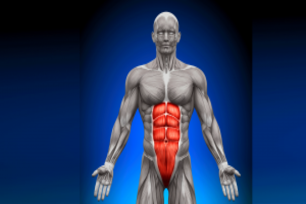
In popular media, the term “core” is often narrowly associated with abdominal muscles, neglecting the critical role played by other passive tissues within the core structure, such as bones, cartilage, and ligaments. To comprehend the core’s full functionality, it’s essential to recognize the broader context of the musculoskeletal system and how it operates as a kinetic chain comprising interconnected bones and joints. This system forms a framework that acts as a system of levers, allowing for the controlled initiation, regulation, and prevention of motion through the neurologically regulated production of muscular torque, which is essentially the force generated by muscles causing joint movement.
The musculoskeletal system functions like a kinetic chain, consisting of rigid bones connected by ligaments at the joints, which act as axes around which muscular and gravitational torques interact. In simpler terms, gravity exerts a downward force on the body or an object (like a barbell or dumbbell), creating resistance. In response, the muscles within the body generate tension, regulated by the nervous system, to counteract gravity and facilitate or prevent motion. This muscular tension provides a stable foundation for powerful dynamic movements of the upper and lower extremities, such as throwing, kicking, or blocking.
The skeletal component of the anatomical core encompasses the bones of the pelvic girdle, which includes the right and left hip bones (os coxae) and the sacrum. The pelvic girdle is linked to the torso through the sacroiliac joints, while the hip joints connect the lower extremities to the pelvic girdle. Therefore, the anatomical core serves as the crucial link through which torque and angular velocity are transmitted from the lower extremities to the upper extremities.
The vertebral column, composed of 33 vertebrae, plays a pivotal role in the core’s structure and function. These vertebrae include 7 cervical, 12 thoracic, 5 lumbar, 5 sacral (fused together), and 4 coccygeal (fused together) segments. Among these, the cervical and lumbar regions exhibit the greatest mobility, primarily due to variations in the orientation of facet joints at the cervicothoracic (C7-T1) and thoracolumbar (T12-L1) junctions. The vertebral column permits various movements, including flexion and extension in the sagittal plane (as seen in abdominal crunches), lateral flexion and reduction in the frontal plane (as in a dumbbell side bend), and rotation in the transverse plane (trunk rotation, as in a medicine ball toss). Core movement terminology often incorporates the terms “lumbar” or “trunk” to indicate the primary region of movement.
When considering facet joints between the vertebrae, approximately 1 to 2 degrees of movement in each plane (sagittal, frontal, and transverse) is possible without encountering passive resistance from ligaments and intervertebral discs. This unrestricted range of movement is termed the “neutral zone.” Maintaining the lumbar spine within this neutral zone during resistance exercises is crucial to prevent excessive stress on passive tissues and facilitate the activation of core muscles. The key to preserving the neutral zone and maximizing spinal stability lies in stiffening the vertebral column through muscular tension. The preservation of spinal stability is especially vital when handling various loads and adopting different postures, such as when performing a back squat with a barbell resting on the shoulders. When the lumbar spine remains in a neutral position, the muscles can provide optimal stabilizing support. In contrast, when the lumbar spine is flexed (outside the neutral zone), the spinal extensor muscles become neurologically inhibited from generating tension, and passive tissues (like cartilage, ligaments, and facet joints) assume a more significant role in stabilization, thereby increasing the risk of injury to these structures.
In isolation, passive tissues have limited capacity to stabilize the spine. For instance, a mechanical model of the lumbar spine indicated that, without muscular support, the spine buckled under a compressive load of just approximately 20 pounds (9 kg). Clearly, this level of support is inadequate for bearing body weight or handling additional loads involved in resistance training, sports activities, or daily tasks. Thus, the activation of core muscles is indispensable for meeting spinal stability requirements across all physical activities.
Anatomical Core (Muscles)
In contrast to passive tissues, muscles within the core provide the necessary torque to initiate movement (concentric muscle actions), control movement (eccentric muscle actions), or prevent movement (isometric muscle actions). While abdominal muscles are often the focal point, several other core muscles contribute to both stabilizing the spine and facilitating dynamic movements. It’s crucial to dispel the misconception that a single core muscle, such as the transversus abdominis, holds paramount importance in spinal stabilization. This misconception originated from research indicating that the transversus abdominis was the first core muscle activated during arm-raising tasks. However, this research was limited to assessing a relatively simple movement task. In more complex movements, core muscle activation patterns vary depending on factors like posture, external loads, and breathing patterns. Consequently, the relative importance of any core muscle can change instantaneously, making it task-specific. Various postures and external loads, influenced by gravity, create resistive loads on the spine, ligaments, facet joints, and discs. To maintain spinal stability, these resistive forces must be counteracted by equal and opposing muscular actions.
Different core muscles possess fibers aligned in varying orientations, which create spinal stability through the simultaneous activation of antagonistic muscles on either side of the trunk while allowing for spinal motion when necessary. Therefore, an effective approach to core muscle development involves a diverse array of exercises encompassing both stabilizing (isometric muscle actions) and dynamic (concentric and eccentric muscle actions) functions.
The functional significance of each core muscle depends on its cross-sectional area, fiber alignment, and its role in instantaneous stabilizing or dynamic functions. For instance, some core muscles like the longissimus and iliocostalis of the erector spinae group span several vertebral segments and possess large moment arms (the distance from a joint to the point of muscle attachment on a bone). This anatomical feature makes them ideally suited for generating significant torque for trunk extension. During exercises like the Romanian deadlift, the longissimus and iliocostalis maintain isometric tension, stabilizing the pelvic girdle in an anterior tilt (forward tilt of the pelvic girdle accompanied by lumbar spine extension). This allows the gluteus maximus and hamstring muscles to dynamically control the alternating extension and flexion movements of the hips. It’s crucial to emphasize the concept of a “hip hinge” when coaching this exercise.
Conversely, other core muscles, such as the rotatores, intertransversalis, and interspinalis, are rich in proprioceptors (e.g., muscle spindles), making them adept at sensing rotation in specific intervertebral facet joints. Their role as position transducers enables the activation of larger superficial muscles to meet spinal stabilization requirements. Furthermore, some core muscles are designed to transfer torque and angular velocity from the trunk to either the lower or upper extremities.
In light of these distinctions, core muscles can be broadly classified into three categories: global core stabilizers, local core stabilizers, and upper and lower extremity core-limb transfer muscles. This section’s intent is to provide a basic overview of some key muscles involved.
Anatomical Core (Neural Integration)
The nervous system plays a central role in orchestrating the complex interplay of core muscles, determining their specific activation patterns and intensities required to stabilize the spine and facilitate the dynamic transfer of torque and angular velocity between different skeletal segments. This neural integration is crucial for enabling efficient and powerful movement patterns in sports and daily activities.
Sports performance is not solely dependent on the absolute production of muscular torque (i.e., strength). If it were, the strongest individuals would dominate sports like baseball and basketball. However, the reality is that even the strongest individuals may not be able to throw a 100-mile-per-hour fastball or excel in other sports that require precise and powerful movements. Absolute muscular torque production is not enough on its own; it must be combined with the neurologically orchestrated steering of torque to optimize the storage and release of muscular elasticity.
Muscles possess an inherent elastic property that allows them to store and release energy, much like a rubber band. This elasticity contributes significantly to the performance of sports skills. However, harnessing this elastic recoil efficiently depends on movement technique rather than pure strength. This is why training methods that focus solely on isolated muscle development often don’t translate into better sports performance. For effective training in dynamic sports, exercises should incorporate ground-based movements that engage multiple muscles, emphasizing both stabilizing (isometric muscle actions) and dynamic (concentric and eccentric muscle actions) functions.
The central nervous system, comprising the brain and spinal cord, continuously receives sensory feedback from proprioceptors, such as muscle spindles, Golgi tendon organs, and free nerve endings. This feedback informs the nervous system about various aspects, including muscle length, tension, joint position, and the rate of joint rotation. Notably, the nervous system must simultaneously address the demands of spinal stability and the need for rhythmic breathing. The rhythmic action of breathing can momentarily compromise spinal stability as core muscles may relax during exhalation. In some instances, such as maximal lifts, lifters adopt the Valsalva maneuver, exhaling against a closed airway, to increase intra-abdominal pressure, enhancing the compressive forces between adjacent vertebrae to maintain spinal stability. However, for most training scenarios involving submaximal torque production, a balance between breathing and core muscle activation is crucial.
Traditionally, exercise instructions have emphasized inhaling during the lowering phase and exhaling during the lifting phase. In reality, breathing during exertion rarely follows such a coordinated pattern. Therefore, coaches should encourage athletes to breathe naturally while maintaining constant tension (abdominal bracing) within the core muscles. As athletes progress from simple to complex resistance exercises, their nervous systems adapt to meet the combined requirements of breathing and spinal stability. The specific combination and intensity of core muscle activation during a given task rely on a combination of feed-forward and feedback mechanisms. Feed-forward mechanisms involve anticipatory muscle activation based on muscle memory from prior experiences. Feedback mechanisms, on the other hand, develop as athletes repeatedly practice and refine sports skills, with the nervous system storing sensory feedback regarding the optimal combination and intensity of core muscle activation required for spinal stability and efficient movement. For example, before a baseball shortstop reacts to field a ground ball, rapid anticipatory core muscle activation occurs to create spinal stability and allow for dynamic hip movement when fielding the ball. Practicing these skills refines sensory feedback, enhancing anticipatory muscle activation during actual game situations.
Proprioceptors found in the intervertebral discs, vertebral ligaments, and facet joint capsules play a pivotal role by relaying sensory feedback to the central nervous system about the position and movement of the vertebral column. This feedback is essential for stimulating specific neural recruitment patterns of the core muscles to meet the demands of a particular task. During any given task, the core muscles must activate sufficiently to stabilize the spine but not excessively to the point of restricting movement. Striking a balance between stiffness and mobility is a critical aspect regulated by the nervous system. Athletes can improve their performance by enhancing the regulation of core muscle activation through proper movement training.
Biomechanics of the Anatomical Core in Sports Performance
From a biomechanical perspective, the core functions as the kinetic link connecting the upper and lower extremities. The skeletal system can be envisioned as a kinetic chain, comprising segments or links connected at joints. Muscles in the body attach to the skeleton through tendons, producing force that is transferred to the skeleton to create torque (muscle force causing joint movement). Therefore, the musculoskeletal system functions as a series of levers generating the necessary torque for initiating, controlling, or preventing movement. The magnitude of muscular torque generated is influenced not only by the force generated by muscles but also by the length of the moment arm relative to the joint axis. Creating sufficient spinal stability through muscular torque depends not only on muscular strength but also on utilizing techniques that leverage the moment arm’s advantage, such as abdominal bracing.
In sports involving ground-based movements, torque generation begins in the lower extremity muscles and progressively increases through sequential activation of core and upper extremity muscles. The timing of muscle activation is crucial to maintain spinal stability and maximize the angular velocity of the involved skeletal segments. In sports requiring throwing movements, achieving maximal angular velocity in the upper arm, through the summation of torque generated by the lower body, core, and upper extremities, enables high-speed release or impact of the object being thrown or struck (e.g., baseball, softball, volleyball, tennis). Similar principles apply to other sports skills involving punching or striking with implements like tennis rackets or baseball bats. Effective sports skill performance necessitates the coordinated activation and relaxation of multiple muscle groups in a precise neural sequence.
Importantly, movement at one skeletal segment of the core can transfer torque and angular velocity to other skeletal segments positioned superiorly or inferiorly. For example, the pelvic girdle connects to the vertebral column through the sacroiliac joints. When an individual’s feet are planted on the ground and the pelvis tilts anteriorly or posteriorly, it results in hyperextension or flexion of the lumbar spine, respectively. This illustrates the concept of a kinetic chain and demonstrates how weakness in muscles acting on one skeletal segment can lead to excessive stress on muscles acting on adjacent segments, potentially leading to compensatory movements and injury.
Proper positioning and stabilization of the anatomical core are fundamental for enabling efficient and powerful movements of the upper and lower extremities. Exercise selection should focus on training core muscles with coordinated joint actions of the upper and lower extremities. For example, coaches can integrate exercises like the single-arm cable chest press performed in a lunge stance