Knee Injuries
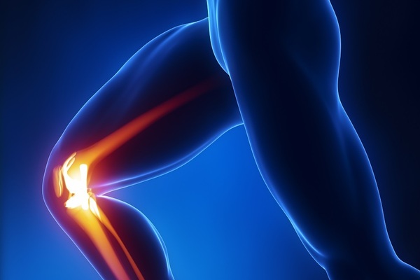
Knee injuries will soon match and likely surpass in incidence the most common injury in the weekend warrior, which is back pain. Injuries to the knee can be debilitating, but thanks to terrific advances in diagnosis and both nonoperative and operative treatments, the athlete can often return to play in a relatively timely manner.
PATELLOFEMORAL PAIN
Common Causes
Patellofemoral pain, also known as anterior knee pain, is the most common complaint of pain in the knee and is most associated with athletic overuse. Patellofemoral pain spans all age groups and all sports and is aggravated by flexed-knee activities such as sitting, climbing stairs, and driving.
Identification
The athlete typically has pain in the anterior knee, but this pain might radiate posteriorly in the case of associated patellofemoral degenerative changes (more common in the older athlete). Significant swelling is unusual, though knee buckling might occur. Localized tenderness might spread over any aspect of the patella. Tenderness at the anterior medial joint line is common. Patellofemoral tracking should be evaluated. The angle formed by the pull of the rectus femoris and patellar tendon is called the quadriceps angle and might be increased in athletes with poor tracking. Ligament tests can help rule out ACL injury. A physical exam should include an overall evaluation of the lower extremity alignment as well as the ipsilateral hip. An X-ray might show patellofemoral malalignment and bony dysphasia as well as degenerative changes.
Treatment
The vast majority of patellofemoral disorders improve with nonoperative treatment. Typically, either a home exercise program or formal physical therapy is recommended. These programs focus on increasing flexibility and strengthening both the quadriceps and hamstring muscle groups. Ultrasound and electrical stimulation might assist in rehabilitation. If swelling or severe pain occurs, nonsteroidal anti-inflammatory medication might be useful. Some athletes might also find an adjustable brace helpful. During rehab, the athlete should avoid locking the knees. Any extreme positions of bending (cross-legged sitting, kneeling, or squatting) or straightening (supporting the leg on a coffee table) should also be avoided. If the patient is not doing well after six months of treatment, MRI can help evaluate the other possible causes of symptoms. Surgical results for patellofemoral pain problems are variable.
Return to Action
Typically athletes refrain from the involved activity for a few weeks up to six months. Athletes who have surgery might take three to six months to recover before returning to sports. Athletes must be able to perform motions mimicking their sport without significant pain before returning. If pain or weakness remains, more therapy is recommended. Patellofemoral braces are available, but these are adjuncts, not remedies.
ILIOTIBIAL BAND SYNDROME
Common Causes
Irritation of the iliotibial band (ITB) as it crosses the lateral aspect of the knee to its insertion on the tibia is often related to overuse activity of the knee and is frequently diagnosed in cyclists, runners, and triathletes. ITB is most commonly associated with increases in training volume.
Identification
The pain is localized to the ITB in the lateral side of the knee; the remainder of the knee might be asymptomatic. The athlete usually experiences the pain at some distance into a training session, making the session difficult or impossible to complete. Typically, little pain occurs at the onset of activity and there is no complaint of swelling. Diagnosis for this injury is the physical exam. Well-localized tenderness spreads over the iliotibial band as it crosses the lateral aspect of the knee. There is typically no localized soft tissue swelling. The ITB might be tight and irritated by front-to-back movement of the band during the flexion–extension movement of the knee. Occasionally, proximal tenderness of the band in the hip area might also occur. The area of lateral knee tenderness should be differentiated from the lateral patella and lateral joint line. Range of motion is normal, and the remainder of the knee exam is normal. Some localized soft tissue irritation might be present in the area of the ITB. Knee X-rays, usually normal, should be run to rule out other injuries.
Treatment
Treatment of ITB syndrome is almost always nonoperative and begins with rest. Supervised physical therapy is very useful in treating this disorder. ITB stretching is difficult for the athlete to perform adequately on his or her own, so it is best to have a physical therapist assist. In addition to stretching the ITB, local modalities such as ultrasound and electrical stimulation to the area of tenderness adjacent to the lateral femoral epicondyle (on the outer side of the knee) are helpful. The athlete might try a 10-day course of anti-inflammatory medications with the proper precautions. A cortisone injection might also be of benefit. Stretching the lower-limb muscles helps to eliminate tension and irritation of the ITB. Ice massage several times a day, using an ice cube directly over the outer border of the knee, should help eliminate symptoms. The athlete should work on maintaining and improving strength in the lower limbs, emphasizing the hip muscle.
Return to Action
This is one disorder in which exercise technique (especially in cyclists) might contribute significantly to the problem. Serious cyclists might want to work with cycling specialists who can examine their biking position and suggest changes to decrease the irritation of the ITB during the cycling motion. For runners, examining the feet and possibly altering the running shoe or adding an orthotic might prove beneficial. Once symptoms have subsided and the athlete has returned to exercise, a continued program of ITB stretching should be incorporated into training. Mending time for an ITB injury is at least 6 weeks and might take up to 12 weeks. When pain is absent or minimal during daily activities, such as stair climbing or pedaling a stationary bicycle, athletes may consider returning to athletic participation. Return must be gradual and may progress as long as there is no pain.
MENISCAL TEAR
Common Causes
Medial meniscal tears occur in every age group. Athletes under 30 who tear their medial meniscus tend to do so in the course of a traumatic injury to the knee; in extreme situations they might be unable to fully extend the knee (known as a locked knee.) In 30- to 60-year-olds, the athlete may not recall any specific injury but report only that pain developed after a particular activeity. Although medial meniscal tears largely outnumber lateral meniscal tears, they are both caused by twisting. Basketball, American football, and soccer have high rates of meniscal injuries.
Identification
Localized pain with a small amount of swelling after activity is the most frequent complaint. Pain is often aggravated by any rotational leg activity or extremes of motion. Athletes might experience positional pain in the knee while sleeping. They often have difficulty returning to their sport of choice but are not disabled by their symptoms (though if a loss of knee extension occurs, this should be immediately addressed). Almost all injuries that result in painful knees make for discomfort or pain in descending stairs. Check for localized tenderness at the involved joint line. The medial meniscus is located in the medial joint line of the knee, and the lateral meniscus is located in the lateral joint line of the knee. (As a reminder, the medial side of the knee is the side closest to the other knee, whereas the lateral side of the knee is the outside of the knee.) Rotary maneuvers such as flexing and extending the knee while externally and internally rotating the limb can help diagnose a meniscus tear if these actions reproduce pain or symptoms. Hearing a click when rotating the knee might indicate a mobile flap of meniscus. Lateral meniscus tears are sometimes associated with a meniscal cyst, which can be felt as a firm, tender soft tissue swelling at the mid-lateral joint line of the knee. Older athletes with coexisting osteoarthritis might have a mild angular deformity at the knee. Standard X-rays are typically normal, though they might show degenerative changes in older athletes. MRI testing is quite accurate in confirming the diagnosis of a meniscal tear.
Treatment
Blood circulation to the menisci is quite limited, so little biologic healing occurs with these injuries. Occasionally athletes do reasonably well with nonoperative treatment, but in most cases surgery is required. During arthroscopic surgery, doctors remove the least amount of meniscal tissue required to solve the problem. The success of arthroscopic partial meniscectomy is quite good in the absence of associated degenerative joint disease. Long-term issues of degenerative joint disease following partial meniscectomy might be related to the amount of meniscus removed, and these effects are minimized with current arthroscopic techniques. Surgery is typically an outpatient procedure with a swift recovery. Athletes often return to full function within six weeks. A home exercise program or supervised physical therapy speeds recovery. Exercise should focus on lower-limb strengthening, with emphasis on hip exercises to reduce stress on the knee joint.
Return to Action
With conservative care (no surgery), about 30 percent of athletes do well. They can return to sports in about 8 to 12 weeks and are limited by occasional pain and instability. Following surgery, athletes often return sooner, sometimes within four weeks. Although some professional athletes have returned sooner (in as little as two weeks), keep in mind that these athletes are rehabilitating five to six days per week for several hours each day. As with many knee injuries, the use of a knee sleeve or soft brace usually depends on the athlete’s desire to wear one. These devices usually offer more proprioceptive support than actual mechanical support. Athletes returning to sport should progress slowly and monitor swelling and pain. If either occurs, decrease the activity level while continuing with a lower-limb strengthening program with emphasis on hip exercises. Hip strength is critical in supporting the entire lower limb. Ice after exercise to reduce swelling and pain.
MEDIAL COLLATERAL LIGAMENT TEAR
Common Causes
The typical cause of a mild to moderate isolated medial collateral ligament (MCL) tear (or sprain) involves either a contact or noncontact injury with a valgus force directed at the knee (placing the knee in a knock-knee” position). These injuries occur in athletes of all ages (particularly those 16 to 50) but are relatively uncommon in older athletes. Injury to the MCL is common in skiers as well as in American football and soccer players. Athletes are at risk when they are positioned with their knees together with a force pushing from the outer side toward the inner side of the knee.
Identification
The athlete often reports hearing a pop or feeling a tearing sensation in the knee. The pain is typically localized to the inner (medial) side of the knee, particularly at the MCL origin on the upper aspect of the knee. Physical examination of the knee with an isolated MCL injury reveals little, if any, swelling. The athlete might have occasional limited range of motion, especially the final 10 degrees of extension, because of pain in the area of the MCL. Localized tenderness occurs along the course of the MCL that might include the medial joint line. It is common that the most significant tenderness is at the MCL insertion on the upper knee. MCL stress testing and a ligamentous exam can verify an MCL diagnosis and help determine if there is an associated anterior cruciate ligament (ACL) tear or other ligament injury. MRI can also verify if the injury is isolated to the MCL or if the ACL is involved. Rotary stress testing at the knee might be painful. These tests are typically positive in athletes with meniscal tears but will also cause pain in those with an MCL sprain.
Treatment
Virtually all isolated MCL injuries can be managed nonoperatively (though combined multiple ligamentous injuries might require surgery). For a low-grade MCL injury, begin active–passive range of motion immediately, with local ice massage, exercise bicycle as tolerated, and exercises to strengthen the quadriceps. Supervised physical therapy should accelerate recovery. No bracing is required, but a medial–lateral sleeve might provide additional comfort. Athletes must make sure to move the knee to regain full range of motion (even when painful) so the knee does not become stiff. They must not sleep with a pillow under the knee because this might hinder the ability to straighten the knee while walking. For more severe isolated MCL injuries, a period of bracing might be necessary to assist in ligament healing. The brace starts at 30 degrees in a hinged locked brace that is gradually unlocked to allow increased motion and physical therapy as healing progresses. Discontinue bracing after four to six weeks. It is uncommon with this injury to have an associated meniscal tear, but if progress remains limited at three months, check for additional tears with MRI. How stiff the knee is at the onset of therapy will depend on the extent of the tear and how long the knee has been braced. Initially, the goal is to regain full range of motion, which may be painful and stiff at the outset. As progress is made, the athlete should complete an exercise program for lower-limb strengthening with emphasis on the hip muscles. When doing inner and outer thigh strength exercises, the athlete should take care not to stress the medial collateral ligament. Ice should be applied after exercise to reduce swelling and pain.
Return to Action
Recovery might take three months. Return to sport depends on restoration of range of motion, strength, and absence of pain. Athletes will typically use a knee sleeve with supports on the medial and lateral sides as they return to activity. They should progress slowly and gradually increase the amount of time spent in their sport. Athletes should master running and cutting drills before beginning full participation.
ANTERIOR CRUCIATE LIGAMENT TEAR
Common Causes
Rupture of the anterior cruciate ligament (ACL) occurs more often in females than in males, from adolescents to older adults. Noncontact injuries to the knee are responsible for most ACL tears. Pivoting and cutting sports (soccer and basketball) are the most common scenarios for noncontact ACL injuries, whereas most direct contact ACL injuries occur in American football. ACL tears can be associated with meniscal tears or collateral ligament injuries.
Identification
Most athletes who have torn their ACL will hear or feel a pop accompanied by pain and, soon after, swelling, though in rare cases little swelling occurs. Rapid swelling in the knee is typically caused by bleeding associated with the injury. The pain might subside quickly after an ACL injury, but this does not mean the tear or strain is healing. An athlete will experience instability with an insecure sensation while pivoting or loading the knee; an occasional sense of hyperextension of the knee is also common. Tenderness often occurs at the lateral joint line. Critical to diagnosis of the ACL tear is the Lachman test, which evaluates the ACL laxity at 30 degrees of knee flexion and includes the uninjured knee for comparison. If the ligament is intact, there will be an endpoint feeling like tensing a string. Absence of this firm sensation typically signals an ACL tear. The medial, lateral, and posterior ligaments are tested as well. Evaluating range of motion is especially important. Standard X-rays are required but seldom reveal much. Occasionally, asmall piece of bone that has pulled off of the lateral aspect of the tibia might show up on X-ray. This indicates an avulsion fracture and is typically associated with an ACL tear. MRI is quite accurate in diagnosing ACL tears. Typically, MRI findings with a torn ACL include bone bruises at the end of the thigh bone, femur, and posterior tibia; swelling; and an abnormal ACL at the femoral attachment. It is not uncommon to have an associated meniscal injury (see p. 210) with an ACL tear, and this is also diagnosed with MRI.
Treatment
Age, occupation, desired activity, sports involvement, and associated injuries to the knee are all taken into consideration when deciding on treatment for an ACL tear. Nonoperative treatment includes supervised physical therapy to restore range of motion, decrease swelling, and restore strength. With return to activities, athletes in more vigorous sports might use a derotational ACL brace. Thanks to recent advances in arthroscopic ACL reconstructive procedures and more rapid postop recovery and return to sports, surgery for ACL tears is a much more attractive option than it once was. In the very young patient with open growth plates, surgery might be delayed until bone maturation, but there is some controversy about this. In the patient older than 60 years, nonoperative treatment is generally recommended but certainly the octagenerian who skis on a regular basis may opt for surgical reconstruction of the ACL. Whereas nonoperative treatment might be considered in any age group for isolated ACL injuries, it is typically less successful in active and athletic patients. Postsurgery, athletes typically return to school or sedentary work within a week. Athletes will use crutches for one to two weeks and begin physical therapy almost immediately. Physical therapy and a strengthening program continue until the injured knee has 90 percent of the strength of the other knee. Training focuses on strengthening the hamstring and quadriceps muscle groups as well as the other lower extremities. The hamstring muscles are particularly important because they add stability to the injured knee. Hamstring contractions pull the tibia backward, which helps counter the inherent ACL instability, which is a forward glide of the tibia. Also, full-knee extension is critical for long-term knee function and should always be a priority in treatment. Clinical results of ACL reconstruction are quite good, with very low reinjury rates. The most common complication is some residual anterior knee pain. Strengthening the hip muscles during therapy is also extremely important in helping to restore stability in the lower limbs and decrease strain on the reconstructed ligament.
Return to Action
With surgery and rehab, athletes can usually return to sport in about six months. Bracing might be initially beneficial upon return. The decision to brace usually depends on athlete preference and whether any instability remains in the rehabilitated knee. Some medical professionals evaluate the post-ACL athlete via a series of functional tests to assess the knee’s strength and stability. Devices such as isokinetic strength testing machines and a series of hopping tests are used. Athletes must progress slowly in resuming sport activity and perform exercises such as running, cutting, twisting, and jumping to mimic the movements of the sport before beginning full participation.
POSTERIOR CRUCIATE LIGAMENT TEAR
Common Causes
Injuries to the posterior cruciate ligament occur when a front-to-back force is directed straight onto the upper tibia. This injury typically occurs during a fall onto a flexed knee, after trauma to the front part of the extended knee, or when the knee is hyperextended. PCL injuries are most common in contact sports such as American football and in cutting and pivoting sports such as basketball, in which knee hyperextension could occur. These injuries are often overlooked and undiagnosed because pain might be the only symptom.
Identification
Posterior cruciate ligament injuries are much less common than ACL injuries. The athlete with an injured PCL complains of local knee pain, might have swelling, and offers little complaint of instability. The injury is debilitating in that it adversely affects an athlete’s ability to run all out, either because of pain or a sense of not being able to trust the knee. Athletes might say, “The knee just doesn’t feel right.” Diagnosis is confirmed via MRI.
Treatment
Most PCL injuries are treated conservatively. Unless associated injuries cause either instability or increased biomechanical stress, surgery is not usually required. However, extensive rehabilitation is necessary. The focus of rehabilitation is strengthening the quadriceps muscles because these muscles add stability to the PCL-deficient knee.
Return to Action
Expect three to six months of rehab with conservative (nonsurgical) treatment of a PCL injury. At least six months are required to rehabilitate the surgically repaired PCL-injured knee. As with the ACL-injured knee, return to athletics is permitted once the knee is relatively pain free, range of motion is good, and the athlete can complete a series of functional knee tests. Bracing is recommended if the athlete wants it or if any knee instability remains. Athletes must progress slowly when returning to sport and perform exercises such as running, cutting, twisting, and jumping to mimic the movements of the sport before beginning full participation.
LATERAL COLLATERAL LIGAMENT TEAR
Common Causes
Injury to the lateral collateral ligament (LCL) usually occurs because of a force directed from the medial (inner side) knee toward the lateral (outer side) knee. This injury can result from a direct blow in contact sports or by a misstep or sharp pivot during pivoting sports.
Identification
Athletes with an injury to the LCL will have local discomfort along the lateral knee. If the athlete sits and flexes the injured knee and then places the foot of the injured knee over the other knee, pain will flare up at the top of the fibula bone. Feel for a tight band of tissue that travels toward the upper knee—this is the LCL. LCL stress testing and a ligamentous exam can verify an LCL diagnosis and help check for an associated anterior cruciate ligament (ACL) tear or other ligament injury. MRI may also be used to determine whether or not the injury is isolated to the LCL. Rotary maneuvers involving stress testing at the knee might be painful. These tests are typically positive in athletes with meniscal tears but also cause pain in those with LCL sprains.
Treatment
Virtually all isolated LCL injuries can be managed nonoperatively (though multiple ligament injuries might require surgery). For a low-grade LCL injury, the athlete should begin active-passive range-of-motion exercises immediately, local ice massage, exercise bicycle as tolerated, and quadriceps-strengthening exercises. Supervised physical therapy should accelerate recovery. No bracing is required, but a mediallateral sleeve can provide some additional comfort. For more severe isolated LCL injuries, bracing might be necessary to assist in ligament healing. The brace starts at 30 degrees in a hinged locked brace that is gradually unlocked to allow increased motion and physical therapy as healing progresses. Discontinue bracing after four to six weeks. If progress remains limited after three months, MRI might be necessary to check for additional injuries. Physical therapy should emphasize lower-limb strengthening and balance exercises that focus on the hip muscles to reduce stress and strain on the knee. Use caution with hip exercises to avoid stressing the outer leg or lateral collateral ligament.
Return to Action
Overall recovery might take three months. Return to sport depends on restoration of range of motion and strength and resolution of pain. Athletes typically use a knee sleeve with supports on the medial and lateral sides as they return to activity. They should progress slowly and perform exercises that mimic their sport before attempting full participation.
PATELLAR TENDINITIS
Common Causes
Painful knee symptoms associated with patellar tendinitis are commonly related to jumping and repetitive running. This condition is often called “jumper’s knee” and is seen most often in basketball players. The injury might also be related to overuse of the knee.
Identification
The hallmark of patellar tendinitis is localized pain in the proximal portion of the patellar tendon near the lower part of the patella. The pain is aggravated by jumping and running and not typically related to any single traumatic event. When pain is severe, athletes might complain of discomfort while stair climbing and sitting. Swelling is uncommon, and the remainder of the knee exam is normal. MRI might reveal changes in the proximal portion of the patellar tendon that are consistent with a partial tear or thickening of the tendon in chronic cases.
Treatment
Most athletes with patellar tendinitis respond to nonoperative treatment. In acute cases, a 10-day course of approved nonsteroidal inflammatory medications (if tolerated) is recommended. The athlete should begin an exercise program emphasizing quadriceps stretching and strengthening. Ice massage might have some benefit. The athlete should avoid all jumping activities. Most athletes will respond to this conservative treatment. Surgical treatment for patellar tendinitis is quite uncommon. On rare occasions, the athlete who has not responded to more than six months of nonoperative treatment and has an abnormal patellar tendon on MRI might consider surgery. Surgical treatment is usually followed by a three- to six-month recovery period. Cortisone shots are not recommended for this injury. Cortisone may hasten degeneration of the patella tendon.
Return to Action
Return time following nonoperative treatment of patellar tendinitis is highly variable. The condition rarely improves before six weeks. Chronic symptoms are not uncommon and often require repetitive treatment. Some athletes report improvement of symptoms and ability to play using a strap at the level of the mid-patellar tendon, similar to the strap used for tennis elbow. The rare athlete who requires surgical treatment might not return to action for six months. Because there is no guarantee for success of this procedure, return time could be much longer.
PATELLA FRACTURE
Common Causes
Fracture of the patella is almost always caused by a direct blow to the knee. This injury can occur in any sport and causes significant disability.
Identification
Fracture of the patella causes immediate pain, swelling, and reduced motion. Weight bearing is difficult. If a patella fracture is suspected, the athlete should be immediately transported to an emergency room; consultation with an orthopedist is encouraged. X-rays should aid diagnosis and provide information on which to base treatment options.
Treatment
If the fracture does not cause significant misalignment of the patella, four to six weeks of immobilization (via bracing or casting) might be recommended. Following this, a progressive course of physical therapy can restore motion, strength, and overall knee function. If surgery is required, the knee is immobilized postoperatively for a shorter time, which allows earlier range-of-motion exercises. Weight bearing should be avoided for about four to six weeks, which could cause atrophy and weakness in the knee. It is thus important to maintain lower-limb strength via straight-leg raises in all positions at least four times a week, adding resistance as tolerated. Begin with resistance above the knee and progress to the ankle as long as no stress is felt on the knee.
Return to Action
Return to sport takes at least eight weeks. The athlete must have good range of motion and strength in the involved knee before attempting a return. Depending on the sport, recovery time might take three to six months, particularly for contact sports.
PATELLOFEMORAL INSTABILITY
Common Causes
Instability of the patellofemoral joint can be caused by either an acute episode or recurrent subluxations or dislocations. The injury might result from trauma such as a direct hit in American football or from a twisting movement in a noncontact sport. Younger athletes often fall victim to patellofemoral instability. An athlete who has been injured this way once must take precautions because this injury tends to recur.
Identification
Dislocation of the patella is a distinct event that incapacitates the athlete. In many cases, the athlete might recall only that something gave out in the knee because most dislocations occur spontaneously with extension of the knee. Following a patellar dislocation, check for localized tenderness in the medial aspect of the knee relating to an injury to the medial patella or medial patella retinaculum. Tenderness is noted on the inner aspect of the knee and is associated with tearing of the medial patellofemoral ligament, an important medial stabilizer of the patella. Significant dislocation of the patella, in which the patella moves out of its groove, may cause a fracture of the patella or of the underlying femur bone; this is known as an osteochondral fracture.
Treatment
Treatment of an acute patellar dislocation is somewhat controversial. Uncommonly, the athlete must be taken to an emergency room for a closed reduction of the patella. Most athletes can be managed nonoperatively with a two-week period of immobilization followed by an aggressive rehabilitation program emphasizing quadriceps and hip muscle strengthening. X-rays might show a small fleck of bone representing an osteochondral fracture with a loose body. MRI may show effusion, tearing of the medial retinaculum or medial patellofemoral ligament, bone contusions, or possible osteochondral fractures of either the medial patellar or lateral femoral condyle. Athletes who have suffered an osteochondral fracture with a loose body or those with significant tearing of the medial restraints of the patella might require surgical intervention. It is very difficult to repair these fractures because very little residual bone remains. Some athletes require repair of the torn medial structure.
Return to Action
Return time varies depending on the degree of soft-tissue damage. A return to sport might require almost three months, and the athlete wears a patellar-aligning brace during initial activity. A postoperative recovery requires a period of bracing and crutches for six weeks followed by extensive rehabilitation, delaying return time at least six months. Surgery tends to have excellent results in preventing further patellar dislocation, but some athletes might have mild residual pain.
OSGOOD-SCHLATTER’S SYNDROME
Common Causes
Osgood-Schlatter’s syndrome is a painful condition affecting growing children. Symptoms tend to be caused by running and jumping activities involving virtually any sport. Pain is related to the growth plate of the tibial tubercle (the bump on the leg just below the knee cap) where the very strong patellar tendon inserts. Once this growth plate closes, symptoms subside. The growth plate closes at a younger age in females than in males.
Identification
Osgood-Schlatter’s is characterized by well-localized tenderness of the tibial tubercle. Look for mild soft tissue swelling, warmth, and prominence of the tubercle. Again, the condition is commonly seen in the growing athlete. Rarely, adults with a history of Osgood-Schlatter’s might continue to have episodic pain at the tibial tubercle when an infused bone ossicle (a small piece of bone attached to the patellar tendon) persists at the tibial tubercle. This can be seen on a lateral X-ray.
Treatment
Virtually all cases of Osgood-Schlatter’s syndrome can be treated nonoperatively. In the past, immobilization was recommended, but it is not required. Treatment is directed toward the degree of painful symptoms. Options include initial cessation of the related activity, local treatment with ice massage and stretching, and strengthening exercises. If symptoms are severe, a seven-day course of an anti-inflammatory medication can be considered. For an adult patient with a symptomatic tibial tubercle with evidence of an unfused ossicle, surgery might be considered to remove the ossicle. The majority of unfused ossicles are asymptomatic. There should be no surgery for cosmetic reasons on an unusually large tibial tubercle.
Return to Action
Return time is dictated by the degree of symptoms and the young athlete’s ability to perform his or her sport. Often they return to sport with mild recurrent symptoms. Again, this condition resolves itself once the growth plate in the maturing athlete closes.
OSTEOCHONDRITIS DISSECANS
Common Causes
Osteochondritis dissecans (OCD) is usually caused by repetitive trauma. A fragment of subchondral bone in the knee joint becomes loose and leads to vague pain and disability. This condition is most common in athletes undergoing repetitive stresses across the knee. Athletes participating in gymnastics and baseball seem most prone to OCD.
Identification
OCD usually occurs below the age of 18 and is twice as common in males as females. The injury is most often seen behind and to the outer side (posterolaterally) of the medial femoral condyle (about 80 percent of cases), and less often seen in the posterior aspect of the lateral femoral condyle. Tenderness occurs along the joint lines, and loading of the joint causes pain. Athletes might sometimes rotate the lower leg when walking to relieve pressure. Pain is most notable during weight bearing and when the involved knee is extended straight. X-rays usually reveal a lesion on the femoral condyle.
Treatment
Because healing of this injury requires as much stress removal as possible, weightbearing restrictions last longer than usual. Conservative treatments might require several months of bearing no weight on the involved knee, requiring extensive use of crutches. If surgical treatment is selected, a period of a few months of reduced weight bearing is still necessary. With the lack of weight bearing and the use of crutches, the leg atrophies and weakens, so athletes must focus on maintaining lower-limb strength via straight-leg raises in all positions at least four times a week, adding resistance as tolerated. Begin with resistance above the knee and progress to the ankle as long as no stress is felt on the knee.
Return to Action
Whether treatment is conservative or operative, recovery time is at least six months. Return to play is advised only after careful reassessment via X-ray and a clinical exam. Range of motion should be pain free in the involved knee.