Functions and Structure of the Skeletal System
The skeletal system, a framework of bones that forms the structural foundation of the human body, is an intricate marvel that serves a multitude of indispensable functions. This article offers an in-depth exploration of the skeletal system, shedding light on its vital roles, the impact of factors like posture, physical activity, and nutrition, and the pivotal interplay between the skeletal and muscular systems.
The Multifaceted Functions of the Skeletal System:
The skeletal system is a dynamic and versatile entity, contributing to various critical functions that are essential for human life and movement:
- Shape and Form: One of its primary functions is to provide the body with its characteristic shape and form. It is the bony framework upon which the entire body is built.
- Support: The skeletal system offers structural support, helping the body to stand upright against the pull of gravity. Without this support, maintaining an upright posture and performing everyday movements would be impossible.
- Protection: Bones act as a protective shield for vital organs. The skull guards the brain, the rib cage shields the heart and lungs, and the vertebral column protects the spinal cord from injury.
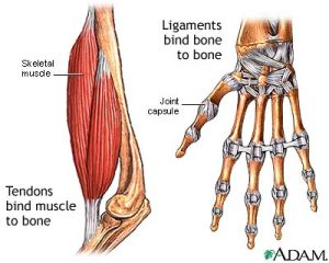
- Facilitation of Movement: The skeletal system plays a central role in bodily movement by serving as an anchor for muscles. Muscles are connected to bones via tendons, and when muscles contract, they generate forces that act on the bones, resulting in movement.
- Blood Production: The interior of certain bones, such as the long bones in the arms and legs, houses bone marrow. This marrow is responsible for producing blood cells, including red blood cells that carry oxygen and white blood cells that help fight infections.
- Mineral Storage: Bones also serve as mineral reservoirs, storing essential minerals like calcium and phosphorus. When needed, these minerals can be released into the bloodstream to maintain proper mineral balance in the body.
The Impact of Posture, Physical Activity, and Nutrition:
The skeletal system’s growth, development, and functionality are profoundly influenced by three key factors: posture, physical activity, and nutrition. Here’s how these factors interplay with the skeletal system:
- Posture: Maintaining proper posture is crucial for the skeletal system’s health and efficient functioning. Poor posture can lead to structural imbalances and musculoskeletal issues over time. Conversely, good posture ensures the effective distribution of forces acting on the body, promoting skeletal well-being.
- Physical Activity: Regular physical activity is vital for skeletal health. Weight-bearing exercises, such as walking, running, and resistance training, help to strengthen bones and maintain bone density. Conversely, a sedentary lifestyle can contribute to conditions like osteoporosis, characterized by weakened bones.
- Nutrition: Nutrition plays a pivotal role in skeletal health. Calcium and vitamin D are essential for bone development and maintenance. A diet lacking in these nutrients can lead to conditions like osteoporosis, which weaken bones and make them more susceptible to fractures.
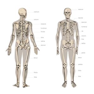
The Axial and Appendicular Skeleton:
The skeletal system is divided into two main divisions: the axial and appendicular skeleton.
- Axial Skeleton: The axial skeleton includes the skull, rib cage, and vertebral column. Comprising approximately 80 bones, it forms the body’s central axis and provides crucial protection for the brain, heart, and spinal cord.
- Appendicular Skeleton: The appendicular skeleton encompasses the upper and lower extremities, as well as the shoulder and pelvic girdles. With roughly 126 bones, it facilitates a wide range of movements and is instrumental in locomotion.
The Role of Joints:
Within the skeletal system, there are over 300 joints where movement occurs. Joints serve as the pivotal points where bones articulate and connect with one another. These junctions are where muscle contractions translate into motion. Joints are critical for fluid and coordinated movement, allowing the body to perform a wide array of activities.
The Dual Functions of Bones:
Bones play two fundamental roles in the context of movement:
- Leverage: Bones act as levers when muscles exert force upon them. This mechanical advantage allows muscles to generate motion efficiently.
- Support: The skeletal system provides structural support for the body, ensuring that it can bear the gravitational forces acting upon it. Proper posture is essential for optimal support and load distribution.
Bones: The Architectural Wonders of the Skeletal System
Bone Growth and Remodeling:
The human skeletal system is a dynamic and ever-changing framework that constantly renews itself through a remarkable process called remodeling. This intricate dance of resorption and formation involves the orchestrated efforts of two specialized cell types: osteoclasts and osteoblasts.
- Resorption: Osteoclasts, the bone-resorbing cells, play a pivotal role in the remodeling process. They break down and remove old bone tissue, a crucial step in maintaining bone health and functionality.
- Formation: On the flip side, osteoblasts, the bone-forming cells, are responsible for laying down new bone tissue. They replace the old, ensuring that the skeletal system remains robust and capable of supporting the body’s functions.
Throughout childhood and adolescence, the pace of bone formation outpaces that of bone removal. This period of growth results in larger, heavier, and denser bones. For most individuals, this process continues until peak bone mass is achieved, typically in their thirties.
It’s important to note that the remodeling process aligns itself with the lines of stress applied to the bone. Consequently, exercise and habitual posture become pivotal influencers of skeletal health. Proper alignment and exercise technique support a positive remodeling process that reinforces healthy posture.
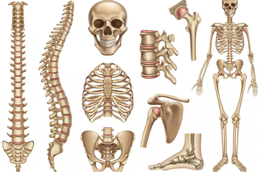
Types of Bones: The Structural Diversity of the Skeletal System
The skeletal system comprises five major types of bones, each uniquely shaped and sized to fulfill specific functions:
- Long Bones: Characterized by their elongated, cylindrical shape with irregular or widened ends, long bones are designed for efficient force distribution. Examples of long bones include the clavicle, humerus, radius, ulna, femur, tibia, and more. Their predominantly compact bone composition ensures strength and stiffness.
- Anatomic Features of a Long Bone:
- Epiphysis: The end of a long bone, primarily composed of cancellous bone and housing red marrow. It’s a critical site for bone growth and may be vulnerable to injury during growth phases.
- Diaphysis: The shaft of a long bone, mainly composed of compact bone, provides support.
- Epiphyseal Plate: The region connecting the diaphysis to the epiphysis, essential for bone length growth during development.
- Periosteum: A fibrous membrane coating the bone, housing nerves, blood vessels, and bone-producing cells. It’s crucial for nutrition, repair, and tendon attachment.
- Medullary Cavity: A hollow space within the diaphysis containing fatty yellow marrow, serving as an energy reserve.
- Articular (Hyaline) Cartilage: A tissue covering the ends of articulating bones, reducing friction in synovial joints.
- Anatomic Features of a Long Bone:
- Short Bones: Short bones exhibit similar length and width, with a somewhat cubical appearance. These bones, including the carpals in the hands and tarsals in the feet, are primarily composed of spongy bone tissue, which maximizes shock absorption.
- Flat Bones: Thin and flat, flat bones feature two layers of compact bone tissue surrounding a layer of spongy bone. They provide protection for internal structures and offer ample attachment sites for muscles. Examples include the sternum, scapulae, ribs, ilium, and cranial bones.
- Irregular Bones: These uniquely shaped bones defy classification into other categories. Vertebrae, pelvic bones, and certain facial bones are among the irregular bones.
- Sesamoid Bones: Small bones embedded in joint capsules or found where tendons pass over joints. They develop within tendons subjected to friction or tension, enhancing leverage and joint protection.
Bone Markings: The Blueprints of Muscular and Tendon Attachments
Most bones possess distinct surface features known as bone markings. These markings come in two primary categories: depressions and processes.
- Depressions: These are flattened or indented areas of the bone, such as fossae and sulci. Fossae, like the supraspinous and infraspinous fossae on the scapulae, serve as attachment sites for muscles. Sulci, like the intertubercular sulcus between the greater and lesser tubercles of the humerus, allow tendons to pass through.
- Processes: Processes are projections that extend from the bone and serve as attachment sites for muscles, tendons, and ligaments. Common processes include the spinous processes of vertebrae, acromion and coracoid processes of the scapulae, and condyles, epicondyles, tubercles, and trochanters found in various bones. These structures play vital roles in skeletal stability, movement, and muscle action.
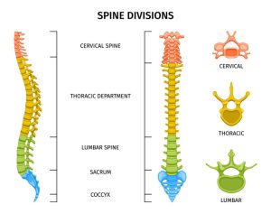
The Vertebral Column: The Backbone of Human Support and Mobility
The vertebral column, also known as the backbone or spinal column, is a critical structural component of the human body, composed of a series of irregularly shaped bones called vertebrae. These vertebrae can be categorized into five distinct regions based on their location and function.
- Cervical Vertebrae (C1-C7): The first seven vertebrae, located at the top of the spinal column, form the cervical spine. These bones provide essential support and mobility for the head, allowing various movements, including nodding and rotation.
- Thoracic Vertebrae (T1-T12): Positioned in the upper and middle back, the twelve thoracic vertebrae are integral components of the thoracic spine. They work in conjunction with the ribs, contributing to the framework of the rib cage. Thoracic vertebrae are larger than cervical vertebrae and increase in size from the top to the bottom.
- Lumbar Vertebrae (L1-L5): The lumbar spine comprises the five lumbar vertebrae, which are the largest in the spinal column. These vertebrae bear the majority of the body’s weight and provide support to the lower back muscles. Due to their significant load-bearing role, the lumbar spine is a common area for discomfort and pain.
- Sacrum: Below the lumbar vertebrae lies the sacrum, a triangular bone formed by the fusion of four or five sacral vertebrae during adulthood. The sacrum connects the vertebral column to the pelvis and serves as a sturdy foundation for the spinal column.
- Coccyx (Tailbone): Situated at the base of the vertebral column, the coccyx, or tailbone, is formed by the fusion of three to five bones in adulthood. It serves as a point of attachment for numerous muscles.
In between each pair of adjacent vertebrae are intervertebral discs composed of fibrous cartilage. These discs act as shock absorbers, cushioning the spine and enabling its flexibility and movement. Intervertebral discs also play a role in maintaining the overall height of the vertebral column.
The vertebral column has several crucial functions beyond merely supporting the body’s structure. It provides attachment points for various muscles, the ribs, and some organs. Additionally, it acts as a protective enclosure for the spinal cord, which controls most bodily functions.
One important consideration in maintaining a healthy vertebral column is achieving the optimal arrangement of its curves, often referred to as a neutral spine. This position minimizes the load and stress on the vertebrae and associated structures. The adult human spine typically exhibits three major curvatures:
- Posterior Cervical Curvature: This refers to the posterior concavity of the cervical spine, which helps balance the weight of the head and facilitates head movements.
- Anterior Thoracic Curvature: The thoracic spine exhibits a posterior convexity, contributing to the chest’s shape and the support of the ribcage.
- Posterior Lumbar Curvature: The lumbar spine features a posterior concavity, allowing it to withstand the substantial forces placed on the lower back.
Overall, the vertebral column is a marvel of anatomical engineering, enabling humans to stand upright, maintain balance, move efficiently, and protect the vital spinal cord. Proper care and posture are essential to ensure the health and functionality of this central structure within the human body.
Joints: The Articulations of the Body’s Framework
Joints, also known as articulations, play a fundamental role in the human body by connecting one bone to another. These structures enable a wide range of movements and provide the body with its mobility. Joints can be classified based on both their structure and their function, with various types of motion occurring during functional movement.
Types of Joint Motion (Arthrokinematics): Joint motion can be categorized into three major types: roll, slide, and spin. It’s essential to recognize that these motions rarely occur in isolation but often combine during functional movements.
- Rolling Motion: In a rolling movement, one joint’s surface rolls across the surface of another, similar to how a bicycle tire moves along the street. An example in the body is the rolling of the femoral condyles over the tibial condyles during activities like squatting.
- Sliding Motion: Sliding movement involves one joint’s surface sliding across another, akin to a bicycle tire skidding across a street. A human example is the tibial condyles sliding across the femoral condyles during knee extension.
- Spinning Motion: In spinning movement, one joint surface rotates on another, similar to twisting the lid off a jar. For instance, the head of the radius (a forearm bone) rotates on the end of the humerus during forearm pronation and supination.
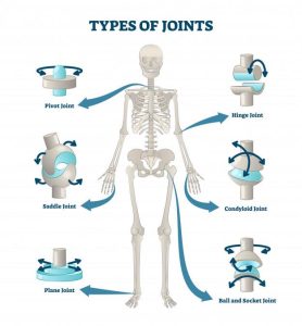
Classification of Joints: Joints in the human body can be broadly categorized into two main types: synovial and nonsynovial joints.
- Synovial Joints: Synovial joints are the most common type of joints in the body, constituting approximately 80% of all joints. These joints are highly mobile and are characterized by the presence of a synovial capsule surrounding the joint, a synovial membrane (the inner layer of the capsule), and hyaline cartilage that cushions the ends of the articulating bones. Synovial joints produce synovial fluid, which lubricates joint surfaces and nourishes the cartilage. There are several types of synovial joints, including:
- Gliding (Plane) Joints: These nonaxial joints allow simple back-and-forth or side-to-side movement. Examples include joints between tarsal bones and carpals.
- Condyloid (Ellipsoidal) Joints: Condyloid joints involve an oval-shaped condyle fitting into an elliptical cavity. These joints permit flexion, extension, and limited abduction, adduction, and circumduction. Examples include wrist joints and metacarpophalangeal joints.
- Hinge Joints: Hinge joints are uniaxial, primarily allowing movement in the sagittal plane. They include joints like the elbow, interphalangeal joints, and the ankle.
- Saddle Joints: Saddle joints resemble a rider in a saddle. They enable movement in two planes—flexion/extension and abduction/adduction—plus circumduction. The thumb’s carpometacarpal joint is an example.
- Pivot Joints: Pivot joints are uniaxial, allowing rotation, pronation, and supination in the transverse plane. Examples include the atlantoaxial joint at the base of the skull and the proximal radioulnar joint at the elbow.
- Ball-and-Socket Joints: These highly mobile joints permit movement in all three planes. Notable examples are the shoulder and hip joints.
- Nonsynovial Joints: Nonsynovial joints lack a joint cavity, fibrous connective tissue, or cartilage in the connecting structure. As a result, they offer minimal to no movement. Examples include the sutures of the skull, the distal tibiofibular joint, and the symphysis pubis (pubic bones).
Joints, with their diverse types and functions, are integral to the body’s ability to perform various movements and maintain structural integrity. Understanding the characteristics of different joint types is crucial for assessing movement, preventing injuries, and promoting overall musculoskeletal health.
The Multifaceted Functions of Joints in the Musculoskeletal System
Joints are pivotal elements of the musculoskeletal system, serving a multitude of essential functions that facilitate movement, maintain stability, and enable coordination throughout the body. Understanding the intricate roles of joints is paramount for personal trainers, as it underpins the comprehension of kinetic chain movement and highlights the interdependence of joints in the human body.
- Facilitating Motion and Movement:
- Primary Function: The primary purpose of joints is to enable motion, thereby facilitating human movement. Without joints, the body would be rigid and incapable of performing various activities, from basic functions like walking to complex actions like sports and exercise.
- Muscle Attachment: Joints play a vital role in anchoring muscles to bones, providing the foundation for muscle contraction. As muscles contract, they exert force on the bones they are connected to via tendons, leading to joint movement.
- Providing Stability:
- Joint Stability: While joints allow for movement, they also provide stability during motion. Stability ensures that movement occurs smoothly and precisely without unwanted or excessive motion in undesired directions.
- Muscle Coordination: Joints work in coordination with muscles to stabilize the body during various activities. For instance, when walking, the joints of the lower limb stabilize to maintain balance and ensure a steady gait.
- Kinetic Chain Movement:
- Interconnected Movement: An essential concept in understanding joint function is that all joints in the human body are interconnected. The movement of one joint directly affects the motion of others. This concept underscores the principle of kinetic chain movement, which has profound implications for how the body functions during activities.
- Examples of Kinetic Chain Movement:
- When rolling the feet inward or outward while standing, observe the associated movement of the knees and hips. Similarly, rotating the hips will impact the position of the knees and feet. This demonstrates that altering the movement of one joint inevitably influences adjacent joints.
- Implications for Personal Trainers:
- Awareness of Kinetic Chain: Personal trainers must be well-versed in the concept of kinetic chain movement to design effective exercise regimens and identify movement dysfunctions.
- Joint Dysfunction: Understanding the interconnectedness of joints allows trainers to recognize that dysfunction in one joint can have cascading effects on neighboring joints. Addressing joint dysfunction and promoting optimal joint health are integral aspects of injury prevention and rehabilitation.
The Comprehensive Role of Joint Connective Tissues: Ligaments
Ligaments, a subset of fibrous connective tissues, are integral components of the musculoskeletal system, playing multifaceted roles in maintaining joint stability, transmitting proprioceptive information to the nervous system, and contributing to the overall health and functionality of the human body. Understanding the intricate nature of ligaments is crucial for professionals in various fields, including physical therapy, sports medicine, and personal training.
- Connective Tissue Essentials:
- Definition: Ligaments are strong, flexible, fibrous connective tissues that serve as the connectors between bones in the human body.
- Structural Composition: Ligaments are primarily composed of collagen, a fibrous protein that imparts strength and durability, and elastin, another protein that provides elasticity and resilience.
- Anchors of Joint Stability:
- Static and Dynamic Stability: Ligaments are pivotal in providing both static and dynamic stability to joints. Static stability refers to the inherent strength of ligaments that prevents excessive joint motion under normal conditions. Dynamic stability is the capacity of ligaments to adapt to changing loads and movements, ensuring controlled motion during activities.
- Joint Support: Ligaments function as stabilizing structures that maintain the alignment and integrity of joints, preventing dislocations and excessive movements that could lead to injury.
- Proprioception and Nervous System Input:
- Proprioceptive Function: Ligaments play a vital role in proprioception, the body’s awareness of its position in space. Sensory receptors within ligaments transmit information to the nervous system about joint position, tension, and movement. This feedback is crucial for coordinating muscle contractions, maintaining balance, and preventing injury.
- Reflexive Responses: Ligament-based proprioceptive feedback triggers reflexive responses that protect joints from potential harm. For example, when a ligament detects excessive stretching or tension, it can initiate muscle contractions to stabilize the joint and prevent further stress.
- Tensile Strength and Load-Bearing Capacity:
- Collagen Arrangement: Collagen fibers within ligaments are aligned parallel to the expected lines of force typically exerted on the ligament. This arrangement grants ligaments exceptional tensile strength, enabling them to resist stretching and withstand substantial loads.
- Load Distribution: Ligaments distribute mechanical loads across the joint, preventing excessive stress on individual structures. This load-sharing function helps prevent joint degeneration and damage.
- Vulnerability and Injury:
- Ligament Sprains: Ligaments are susceptible to injury, often manifesting as ligament sprains. These injuries occur when ligaments are subjected to excessive forces or abnormal movements that exceed their tensile strength. Ligament sprains vary in severity, from mild stretching to complete tears, with corresponding implications for joint stability.
- Rehabilitation and Strengthening: Effective rehabilitation and strengthening programs focus on restoring ligament integrity, proprioceptive function, and joint stability. These programs often incorporate exercises to enhance neuromuscular control and promote collagen remodeling in ligaments.
- Clinical and Training Significance:
- Sports Medicine: In sports medicine, understanding ligament function is essential for diagnosing and managing injuries such as ligament sprains or tears. Treatment may involve rest, physical therapy, bracing, or surgical intervention.
- Physical Training: For personal trainers and strength coaches, knowledge of ligament function guides the design of exercise programs that improve joint stability, enhance proprioception, and reduce injury risk. Targeted exercises can strengthen the supportive musculature surrounding ligaments, further bolstering joint stability.
The Impact of Exercise on Bone Mass: Building Stronger Bones through Physical Activity
The human skeletal system, primarily comprised of bones, forms the structural framework of our bodies, providing support, protection, and the means for bodily movement. The health and strength of our bones are influenced by a variety of factors, with exercise playing a pivotal role in enhancing bone mass, density, and overall skeletal health. Understanding the intricate relationship between exercise and bones is essential for individuals of all ages, as it contributes not only to improved bone health but also to enhanced muscle strength, coordination, balance, and the prevention of fractures, especially among older adults and those diagnosed with osteoporosis.
- Bones as Living Tissues:
- Continuous Remodeling: Bones are dynamic and living tissues that undergo a constant process of remodeling throughout life. This process involves two essential phases: resorption, where old bone tissue is broken down and removed by specialized cells known as osteoclasts, and formation, where new bone tissue is generated by osteoblasts to replace the old. In childhood and adolescence, the formation phase typically outpaces resorption, resulting in larger, denser bones.
- Peak Bone Mass: Peak bone mass, characterized by maximal bone density and strength, is usually achieved in one’s thirties. The amount of bone mass accrued by this stage significantly influences one’s susceptibility to osteoporosis later in life.
- Exercise-Induced Bone Benefits:
- Enhancing Bone Strength: Regular exercise, particularly weight-bearing activities, stimulates bones to become stronger and denser. Weight-bearing exercise imposes mechanical forces on bones, effectively challenging them to adapt and grow denser in response to these stresses. This phenomenon is referred to as the “Wolff’s Law,” which states that bones adapt to the loads under which they are placed.
- Prevention of Osteoporosis: Consistent exercise helps maintain and increase bone mass, reducing the risk of osteoporosis, a condition characterized by fragile, porous bones that are highly susceptible to fractures. Weight-bearing exercises, in particular, enhance bone density, especially in the spine and hips.
- Coordination and Balance: Beyond its direct effect on bones, exercise plays a crucial role in enhancing overall coordination and balance, reducing the likelihood of falls and fractures, particularly among older adults.
- Weight-Bearing Exercises:
- Effective Bone Strengthening: Weight-bearing exercises, such as resistance training, walking, bodyweight squats, push-ups, jogging, climbing stairs, and dancing, are highly effective in strengthening bones. These activities require the skeletal system to work against gravity, prompting bone adaptation and improved density.
- Comparison with Non-Weight-Bearing Activities: While activities like swimming and cycling offer numerous health benefits, including cardiovascular fitness and weight management, they are not the most effective means of stimulating bone growth. These non-weight-bearing exercises do not impose the same gravitational forces on bones, limiting their impact on bone density.
- Strategies for Optimizing Bone Health through Exercise:
- Variation in Exercise: To maximize bone health, it’s advisable to incorporate a variety of weight-bearing exercises into one’s fitness routine. This variation ensures that different bones and muscle groups are appropriately challenged.
- Progressive Resistance: Gradually increasing the intensity and resistance of weight-bearing exercises over time is key to continued bone adaptation. This can be achieved by adding more weight, repetitions, or complexity to workouts.
- Balanced Approach: Combining weight-bearing exercises with activities that promote cardiovascular fitness, flexibility, and muscular strength provides a holistic approach to overall health and well-being.
- Lifelong Investment in Bone Health:
- Importance of Early Intervention: Building strong bones during childhood and adolescence is vital, as it sets the foundation for peak bone mass. Encouraging physical activity and a balanced diet rich in calcium and vitamin D in these formative years is essential.
- Continued Benefits in Adulthood: It is never too late to invest in bone health through exercise. Even in adulthood and later stages of life, consistent physical activity can help maintain bone mass, reduce bone loss, and enhance overall musculoskeletal function.