
Upper Extremity
The shoulder joint is a complex and highly mobile joint that plays a crucial role in upper body movement and function. Understanding the movements and muscles involved in the shoulder joint is essential for designing effective exercise programs, improving athletic performance, and preventing injuries. Here, we will expand on the anatomy and movements of the shoulder joint.
Anatomy of the Shoulder Joint: The shoulder joint, also known as the glenohumeral joint, is formed by the articulation between the humerus bone of the upper arm and the scapula bone (shoulder blade). This ball-and-socket joint provides the shoulder with a wide range of motion but also makes it susceptible to instability and injury.
Movements at the Shoulder Joint: Six main movements occur at the shoulder joint, allowing for the versatility and flexibility of the upper limb:
- Flexion: During shoulder flexion, the upper arm is elevated forward, moving toward the face. This movement is commonly seen in activities like reaching overhead.
- Extension: Shoulder extension involves moving the arm backward, behind the plane of the body. It is essential for actions like pulling the arm backward.
- Abduction: Abduction of the shoulder occurs when the arm moves up and out to the side of the body, away from the midline. This movement is seen when raising the arm out to the side.
- Adduction: Adduction is the opposite of abduction. It involves pulling the arm in toward the side of the body, bringing it closer to the midline.
- Internal Rotation: Internal rotation is the movement of the arm inward or medially, as when reaching behind the back or tucking in a shirt.
- External Rotation: External rotation is the opposite of internal rotation, where the arm moves outward or laterally. This movement is often seen when opening a door.
Muscles of the Shoulder: Several muscles contribute to these movements and provide stability to the shoulder joint. The key muscles include:
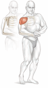
- Deltoid Muscle: The deltoid muscle is the primary muscle responsible for the overall shape of the shoulder. It consists of three separate sections or heads, each capable of moving the arm in different directions:
- Anterior Deltoid: This portion, located in front, attaches to the clavicle and is responsible for raising the arm forward (shoulder flexion).
- Lateral Deltoid: Situated at the side, it attaches to the acromion and is responsible for lifting the arm out to the side (shoulder abduction).
- Posterior Deltoid: Found at the back, it attaches to the scapula and plays a role in moving the arm backward (shoulder extension).
- Rotator Cuff Muscles: The rotator cuff is a group of four muscles that form a protective sleeve around the shoulder joint. These muscles are crucial for shoulder stability and strength. The four rotator cuff muscles are:
- Supraspinatus: Located above the shoulder joint, it raises (abducts) the arm up and outward, as in hailing a taxi.
- Infraspinatus and Teres Minor: These muscles are positioned behind the shoulder joint and are responsible for rotating the arm outward, as in a hitchhiking gesture.
- Subscapularis: Situated in front of the shoulder joint, it rotates the arm inward, such as when folding the arms across the chest.
Understanding the anatomy and movements of the shoulder joint is vital for creating well-rounded exercise programs, improving athletic performance, and addressing shoulder-related issues effectively. Incorporating exercises that target these muscles and movements can help maintain shoulder health and function.
Chest
The chest, or pectoral region, plays a crucial role in upper body movement and strength. Understanding the anatomy and functions of the muscles in this area is essential for designing effective exercise programs, improving athletic performance, and enhancing overall chest development. Here, we will expand on the anatomy and functions of the chest muscles.
Anatomy of the Chest Muscles: The primary muscle of the chest is the pectoralis major. This fan-shaped muscle consists of two distinct heads with different points of origin:
- Upper Clavicular Head: This portion arises from the clavicle (collarbone).
- Lower Sternal Head: It originates from the sternum (breastbone). These two heads merge into a single tendon that attaches to the humerus bone of the upper arm. Notably, as the muscle inserts, the tendon twists so that the upper head attaches beneath the lower head.
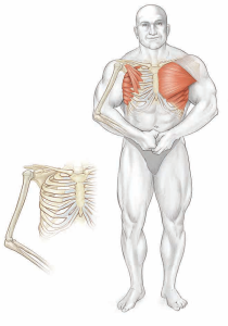
Functions of the Pectoralis Major: The pectoralis major muscle plays a vital role in various movements at the shoulder joint. Depending on the angle through which the arm is moved, functionally, it may be considered as having three sections—upper, middle, and lower. Here are its primary functions:
- Adduction: The pectoralis major contributes to adduction, which involves moving the arm across the chest. For instance, during a push-up or a bear hug, this muscle contracts to bring the arm forward and across the chest.
- Flexion: It assists in flexing the arm at the shoulder joint, allowing for movements such as raising the arm forward.
- Internal Rotation: The pectoralis major aids in internal rotation of the arm, particularly when the arm is flexed, as in throwing a baseball.
It’s important to note that the function of specific fibers within the pectoralis major muscle varies based on the position of the shoulder joint. Some fibers have a better mechanical advantage for creating motion, while others are still active but cannot contract as effectively due to shoulder positioning.
Other Muscles in the Chest Region:
- Serratus Anterior: This muscle forms the side wall of the chest and plays a crucial role in shoulder stability and movement. It arises from behind the scapula (shoulder blade) and wraps around the chest wall to attach to the upper eight ribs. The serratus anterior assists in protracting (pulling forward) the scapula, helping stabilize it against the rib cage. It is particularly active during most chest exercises and works vigorously during the lockout phase of movements like push-ups or bench presses.
- Pectoralis Minor: Situated beneath the pectoralis major, the pectoralis minor muscle has a minor function and does not significantly contribute to the size of the chest. Its role is relatively limited compared to the pectoralis major.
Understanding the anatomy and functions of these chest muscles is essential for targeting them effectively during exercise and ensuring balanced chest development. Incorporating a variety of chest exercises can help individuals achieve their fitness goals and enhance their upper body strength and aesthetics.
Arms
Anatomy of the Arm and Forearm: The arm is a complex structure divided into two main sections: the upper arm and the lower arm (forearm). The upper arm consists of a single bone, the humerus, while the forearm is composed of two bones, the radius (located on the thumb side) and the ulna (located on the little-finger side). The elbow serves as a hinge joint, formed at the junction of the humerus, radius, and ulna. At the elbow joint, two primary movements occur: flexion and extension. During elbow flexion, the forearm moves toward the upper arm, while extension involves the forearm moving away from the upper arm.
Additionally, movement occurs within the forearm due to the rotation of the radius around the ulna. This rotation allows for two essential movements: supination (palm up) and pronation (palm down). These movements take place at the radioulnar joints. The wrist joint marks the junction between the lower end of the forearm bones and the small bones in the hand.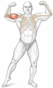
Biceps Brachii: The biceps brachii, aptly named for its two heads, plays a significant role in arm movement. The two heads are as follows:
- Short Head: This head attaches to the coracoid process.
- Long Head: The long head arises from above the glenoid of the shoulder joint. Both heads merge and attach approximately 1.5 inches (4 cm) below the elbow joint onto a tuberosity on the inner side of the radius bone.
Functions of the biceps brachii include:
- Elbow Flexion: It is responsible for bending the elbow joint, allowing actions such as raising the hand toward the face.
- Forearm Supination: The biceps brachii contributes to rotating the forearm so that the palm faces up.
In addition to the biceps brachii, two other muscles play a role in elbow flexion:
- Brachialis: This muscle lies beneath the biceps brachii and attaches to the ulna bone just below the elbow joint. It assists in elbow flexion.
- Brachioradialis: Arising from the outer aspect of the lower end of the humerus, the brachioradialis travels down the forearm to attach to the radius, just above the wrist joint. It contributes to elbow flexion as well.
Triceps Brachii: The triceps brachii is a powerful muscle with three heads (sections):
- Long Head: Arises from beneath the glenoid fossa of the shoulder joint.
- Lateral (Outer) Head: Arises from the outer surface of the humerus.
- Medial (Inner) Head: Arises from the medial and rear surfaces of the humerus.
All three heads merge at their lower ends to form a single tendon, which attaches behind the elbow joint onto the olecranon process of the ulna bone. The primary function of the triceps brachii is elbow extension, which involves moving the hand away from the face. Unlike the three muscles that bend the elbow (biceps brachii, brachialis, and brachioradialis), the triceps brachii is the sole muscle responsible for straightening the elbow joint.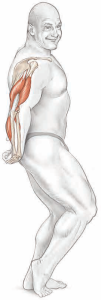
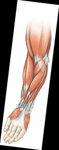
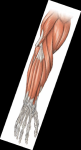
Forearm Muscles: The forearm is a complex region comprising 20 muscles organized into two compartments: the flexor group (palm side) and the extensor group (reverse side). These muscles are involved in various wrist and finger movements. Notably, the wrist flexors and extensors are superficial muscles, while the finger flexors and extensors are deep muscles closer to the bone.
Superficial Wrist Flexors: These muscles cross both the wrist and elbow joints and are involved in wrist movement. They include:
- Palmaris Longus
- Flexor Carpi Radialis
- Flexor Carpi Ulnaris
Superficial Wrist Extensors: These muscles also cross both the wrist and elbow joints, contributing to wrist extension. They include:
- Extensor Carpi Radialis Longus
- Extensor Carpi Radialis Brevis
- Extensor Carpi Ulnaris
Deep Finger Flexors: Responsible for finger movements, including flexion, they include:
- Flexor Digitorum Superficialis
- Flexor Digitorum Profundus
- Flexor Pollicis Longus
Deep Finger Extensors: These muscles enable finger extension and include:
- Extensor Digitorum
- Extensor Pollicis Longus
- Extensor Pollicis Brevis
- Extensor Indicis
In addition, forearm muscles like the supinator and pronator muscles facilitate supination (palm up) and pronation (palm down) movements of the hand, respectively.
Understanding the anatomy and functions of these arm and forearm muscles is crucial for performing a wide range of upper body exercises, enhancing strength, and promoting optimal joint mobility.